• A SURGICAL FEAT ACROSS SYSTEMS: TACKLING RENAL CELL CARCINOMA WITH INTRACARDIAC EXTENSION

    ||A Rare and High-Risk Multidisciplinary Triumph at Rela Institute||A 47-year-old man presented to Rela Institute with a complex and life-threatening condition—clear cell renal cell carcinoma (RCC) that had extended beyond the kidney to invade the liver, adrenal gland, and, most critically, the right atrium of the heart via the inferior vena cava (IVC). What began as persistent cough and breathlessness led to a startling discovery—a massive retroperitoneal tumor with intracardiac extension, a rare presentation seen in less than 1% of RCC cases. Imaging including PET-CT, MRI, and CT-pulmonary angiography revealed a large necrotic mass invading multiple organs and extending into

  • LUNG TRANSPLANT IN A CASE OF INTERSTITIAL LUNG DISEASE

    ||LUNG TRANSPLANT IN A CASE OF INTERSTITIAL LUNG DISEASE|| 42 year old female a known case of interstitial lung disease since 2010. She is also a known case of rheumatoid arthritis. She had worsening of her lung functions since 2020 and became oxygen dependent since 2023. She required more than 10 litres of oxygen support and became totally bedridden for the past 3 months. She got admitted and evaluated for lung transplantation and got registered in the Tamilnadu state transplant registry. She got deteriorated requiring HFNC support with 100 % oxygen. It was found that maintaining oxygen saturation more than

  • LUNG TRANSPLANT IN SEVERE PULMONARY HYPERTENSION

    ||LUNG TRANSPLANT IN SEVERE PULMONARY HYPERTENSION||19-year-old female was diagnosed with Bronchiectasis at 7 years of age. Her lung condition gradually deteriorated which led her to become oxygen dependent. She became extremely oxygen dependent and had breathlessness even at rest. She hardly maintains oxygen saturation more than 85% even with 10 litres of oxygen support. Her family was extremely worried about her declining health condition. Her parents brought her to Rela institute. She was evaluated and was found to have cystic bronchiectasis with severe pulmonary hypertension due to long standing poor pulmonary reserve. Parents were counselled about high-risk lung transplantation and

  • When the Heart Follows the Liver

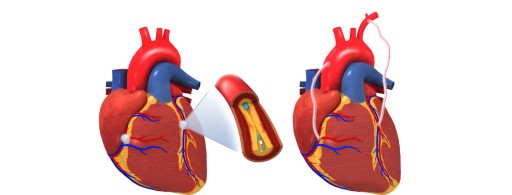
    When the Heart Follows the LiverManaging Cardiac Surgeries in Hepatic CompromiseHIGH-RISK CABG IN A PATIENT WITH LIVER DYSFUNCTIONA 55-year-old gentleman with autoimmune hepatitis, chronic kidney disease, and previous CVA presented with rest angina. Coronary angiogram revealed triple vessel disease. Despite ascites and pulmonary hypertension, he underwent successful high-risk CABG performed by Dr. Srinath Vijayasekharan & Dr Senthil Kumar (Cardiothoracic and Heart & Lung Transplantation Surgeon). Intraoperatively, ischemic MR was noted but managed medically, and mitral valve replacement was avoided. The patient recovered without complications, reflecting a tailored approach to high-risk cardiac cases

  • CABG POST-LIVER TRANSPLANT: A COORDINATED EFFORT

    ||CABG POST-LIVER TRANSPLANT: A COORDINATED EFFORT||Just months after liver transplantation, a 64-year-old man presented with chest pain. Coronary angiogram showed triple vessel disease. He underwent elective off-pump CABG performed by Dr. Srinath Vijayasekharan & Dr Senthil Kumar (Cardiothoracic and Heart & Lung Transplantation Surgeon), which he tolerated well despite moderate PAH. The case show cases how post-transplant cardiac care can be managed successfully through careful planning and multidisciplinary coordination.

  • HEART TRANSPLANTATION IN A CASE OF DILATED CARDIOMYOPATHY

    HEART TRANSPLANTATION IN A CASE OF DILATED CARDIOMYOPATHY 20 year old male presented with complaints of breathlessness (NYHA class III) for the past one year. He is a known case of dilated cardiomyopathy with the history of sudden cardiac death of his sister at a young age. His echo revealed severe global hypokinesia of LV with LVEF of 15%. His 6 min walk test was low (150 meters). His right heart hemodynamic study showed a cardiac output of 3.4 lit and PVR of 9.4 wood units. Inspite of maximum medical management, his symptoms worsened and advised to undergo heart transplantation.

  • AORTIC STENOSIS WITH SEVERE LV DYSFUNCTION

    ||AORTIC STENOSIS WITH SEVERE LV DYSFUNCTION|| A 20-year-old gentleman a known case of bicuspid aortic valve from childhood. He underwent aortic balloon valvotomy in 2017. In last three months he had multiple hospital admissions for breathlessness. On evaluation the echo revealed severe aortic stenosis with severe LV dysfunction (LVEF -25%). He was referred from Tirupathi for further evaluation. In view of severe LV dysfunction, the risks and the need for mechanical circulatory support was explained and the patient was taken up for high-risk aortic valve replacement performed by Dr. Srinath Vijayasekharan, Dr. Senthil Kumar and Team. Intraoperative TEE revealed bicuspid

  • AORTIC ROOT ENLARGEMENT

    AORTIC ROOT ENLARGEMENT 60 years old female presented with complaints of breathlessness (NYHA class III). She was evaluated in outside hospital and was found to have severe aortic stenosis. Patient came here for aortic valve replacement. Intraoperatively TEE revealed severe aortic stenosis with moderate aortic regurgitation. Aortic annulus was sized but it was very small not even admitting 17 mm valve so aortic root enlargement was done between LCC and NCC and augmented with Dacron patch and pericardium. The aortic valve was replaced with 19 mm TTK Chitra aortic valve. Immediate post procedure TEE showed a mean gradient across prosthetic

  • VENTRICULAR SEPTAL RUPTURE

    ||VENTRICULAR SEPTAL RUPTURE|| 51 years old gentleman presented to ER with complaints of breathlessness. He is a known case of CAD and underwent PTCA to LAD and LMCA 15 days back. He was stabilized in ICU and was found to have cardiac murmur for which Echo was done which revealed ventricular septal rupture (VSR). Emergency VSR closure was done by Dr. Srinath Vijayasekharan, Dr. Senthil Kumar and Team, after explaining the risks and complications. Intraoperative TEE revealed 5mm VSR with left to right shunt and moderate LV dysfunction (LVEF – 35%). Bovine pericardial patch closure of VSR and CABG was

Go Back Top